High Content Imaging Core
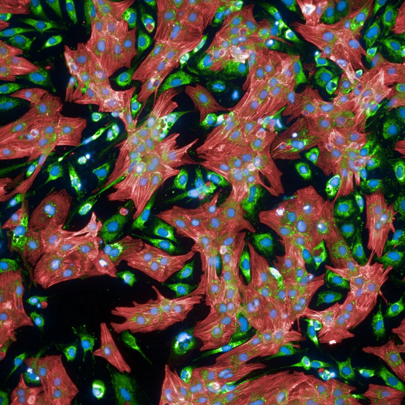
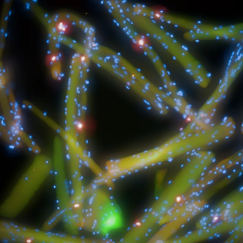
OHRI’s High Content Imaging Core offers training and full access (24/7) to cutting-edge multi-channel fluorescent 3D imaging systems and analysis software.
On this page:
While designed for a fast and fully automated acquisition required in large-scale drug screens, they are also useful for detailed scanning of massive areas such as tissue sections. Not only is it much faster than a traditional microscope, but the fully automated analysis software also allows for completely unbiased object identification and quantification of any biologically relevant data, including cell morphology, signal intensity and kinetic changes.
The systems are suitable for live or fixed cells or tissues in any format of rectangular cell culture plate or slides, but they are not suitable for Petri dishes.
We offer training and access to two different systems: Operetta CLS Live and Arrayscan VTI. Both are equipped with confocal modules and live cell chambers. You can find detailed specifications about each system and its applications in the sections below.
Our core facility offers full training, 24/7 access, experimental support as well as technical support.


Arrayscan VTI
The ArrayScan is a high content imaging instrument that offers a wide set of functions for the high and medium throughput study of cell biology in a single modular platform. From the development of high-content assay, through basic cell biology research to systems biology and drug discovery and toxicology, this system has been engineered to deliver robust data with minimal effort and with the fastest “image-to-answer” currently available.
The high content imaging core is composed of two separate ArrayScan VTI systems, both equipped with an Orbitor RS, capable of processing of up to 100 plates (fully automated). Each system has their own specific modules (live cell chamber, confocal unit, apotome, etc), enabling a wide variety of applications. With a 7-channel LED light engine and the HCS Studio software, the ArrayScan VTI allows the creation of personalized analyses suited for any experimental design.
Operetta CLS Live
The Operetta is a high-throughput microplate imager for high-content analysis (HCA). It can acquire, analyze and manage fluorescence, brightfield and digital phase contrast images. The combination of high-power LED excitation with fast, precise mechanics and one large format sCMOS camera enables fast imaging. Controlled by Harmony software, it offers a seamless workflow for reliable discrimination of phenotypes even in complex cellular models.
The High Content Imaging Core possesses an Operetta CLS Live system, equipped with a live cell chamber, a confocal unit and fully automated water immersion objectives. With an 8-LED kit and the Harmony software, the Operetta CLS Live allows the creation of fully customized analyses suited for any experimental design.
Pricing
$40 per attendee
$20/hour
$60/hour
Time spent on system set-up or gathering data and images from previously executed scans is free.
*Training is required to gain access to the Operetta and the Arrayscan as well as their booking calendars.
Contact us
Dr. Julien Yockell-Lelievre
(613) 737-8899 x79178
Dr. William Stanford’s laboratory (W5214)
Sprott Centre, 5th floor
Ottawa Hospital Research Institute