
Antonio Colavita
PhD
Senior Scientist, Neuroscience
Ottawa Hospital Research Institute
Bio
Dr. Colavita completed a Ph.D. at the University of Toronto and postdoctoral research at Stanford University.
Research Goals and Interests
My laboratory uses the microscopic nematode C. elegans to investigate how neuronal morphology is maintained in mature and aging animals, and to explore early ventral nerve cord morphogenesis, a system that provides insight into how convergent extension and cell-sorting movements shape developing tissues. Understanding how neurons maintain their morphology and how neural tissues undergo morphogenesis is fundamental to explaining why these processes break down in neurodevelopmental and age-related disorders.
 C. elegans is an ideal model for this work: its small size (~1 mm), transparent body, and short generation time make it highly accessible for in vivo imaging and genetic analysis, and its simple anatomy of 959 somatic cells is fully mapped at single-cell resolution. Of these, 302 are neurons with fully mapped axonal trajectories and synaptic connections. C. elegans also offers a powerful genetic toolkit for precise molecular studies. Despite its simplicity, it relies on many of the same pathways used in more complex nervous systems, allowing us to uncover broadly relevant principles of neural development and maintenance.
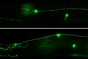
![]() Genetic and molecular mechanisms that establish and maintain neuronal structure. Neurons are among the most morphologically complex cell types, and although their diverse axonal, dendritic, and synaptic structures arise through actin- and membrane-driven remodeling during development, the mechanisms that maintain these architectures in mature and aging animals remain poorly understood. To investigate these mechanisms, we use the six VC motor neurons that regulate egg laying as a model system. These neurons are well suited for studying morphology because they display distinctive bipolar shapes with long, branched axons that extend either along the anterior–posterior (AP) axis or, in the case of the vulval-proximal VC4 and VC5, laterally around the vulva. A major focus of our work is uncovering how these AP and lateral trajectories are formed and maintained.
Genetic and molecular mechanisms that establish and maintain neuronal structure. Neurons are among the most morphologically complex cell types, and although their diverse axonal, dendritic, and synaptic structures arise through actin- and membrane-driven remodeling during development, the mechanisms that maintain these architectures in mature and aging animals remain poorly understood. To investigate these mechanisms, we use the six VC motor neurons that regulate egg laying as a model system. These neurons are well suited for studying morphology because they display distinctive bipolar shapes with long, branched axons that extend either along the anterior–posterior (AP) axis or, in the case of the vulval-proximal VC4 and VC5, laterally around the vulva. A major focus of our work is uncovering how these AP and lateral trajectories are formed and maintained.
The C. elegans ventral nerve cord (VNC): a model for central nerve cord development. Initially, it may seem surprising that VNC assembly in microscopic nematodes and neural tube development in vertebrates share fundamental features. However, we have demonstrated that these processes rely on conserved molecular and cellular mechanisms. Both systems use convergent extension to narrow tissue along the medial-lateral axis while extending it along the AP axis, and both exhibit multicellular rosette assembly and resolution, polarised enrichment of planar cell polarity components such as VANG at cell membranes, and tissue remodeling driven by actomyosin-mediated junctional contractions. With its anatomical simplicity and genetic accessibility, the VNC therefore provides a powerful model for uncovering shared molecular and cellular principles.
 Motor neuron organisation in the VNC as a model for cell sorting. Cell sorting is essential for tissue patterning and morphogenesis, enabling cells to recognise one another, rearrange neighbours, and segregate into distinct spatial domains. At hatching, the VNC contains 6 DD, 9 DA, and 7 DB motor neurons arranged in a stereotyped pattern that emerges from coordinated progenitor movements, including cell-sorting, during the bean to 2-fold stages of embryogenesis. Unlike most cell-sorting studies, which examine mixing between layers or boundary formation, VNC assembly involves the sorting of three distinct neuron types into a higher-order structure that can be tracked in vivo at single-cell resolution. Because each DD, DA, and DB class can be uniquely labelled with fluorescent markers, embryonic sorting defects are readily visualised as mispositioned neurons at the first larval stage, providing an accessible and quantifiable readout.

Figure 1. Time-lapse imaging (80 minutes) of DD and DA neuronal progenitor movements during ventral nerve cord (VNC) assembly (timelapse: J. Evans).

Figure 2. The vang-1/PCP and sax-3/Robo pathways organise motor neuron arrangement in the VNC and act redundantly to regulate the convergent extension movements that drive VNC assembly. Convergent extension defects in vang-1 sax-3 double mutants manifest as anteriorly displaced motor neuron cell bodies in embryonic (A and B) and larval worms (C and D). Single mutants (E and F) display sorting errors.
News

Publications
VNC-Dist: A machine learning-based semi-automated pipeline for quantification of neuronal position in the C. elegans ventral nerve cord
2025-08-28 Go to publicationNuclear hormone receptor regulation of PAL-1/Caudal mediates ventral nerve cord assembly in C. elegans
2025-01-01IPPK-1 and IP6 contribute to ventral nerve cord assembly in C. elegans
2025-01-01Notch-mediated regulation of β-Catenin-TCF activity instructs anteroposterior neuron positioning in C. elegans
2025-01-01VNC-Dist: A machine learning-based semi-automated pipeline for quantification of neuronal positioning in the C. elegans ventral nerve cord
2024-11-18 Go to publication